The human brain is an incredibly complex organ, composed of billions of nerve cells (neurons) and support cells (glial cells), all working together to control thought, movement, emotions, and vital bodily functions.
It contains specialized tissues, fluids, and chemical messengers that allow it to process incoming information and communicate seamlessly with the rest of the body.
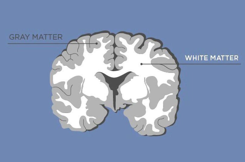
Think of your brain as a highly advanced supercomputer:
Now imagine what would happen if a computer’s wiring were damaged. Information would be delayed, misrouted, or lost entirely leading to errors, system crashes, or poor performance. This is exactly what occurs in a traumatic brain injury (TBI) when either the cerebral cortex or the white matter is disrupted.
In all these scenarios, external forces or environmental stressors disrupt the brain’s normal functioning making timely evaluation and diagnosis essential, even in the absence of a visible head injury.
Many traumatic brain injuries especially those from car accidents, falls, or high-impact sports result in coup-contrecoup and rotational injuries, which often lead to axonal shearing, also known as Diffuse Axonal Injury (DAI). Understanding these mechanisms is essential for proper diagnosis and treatment.
Axonal shearing occurs when the brain is rapidly accelerated or decelerated frequently without direct impact to the skull. These forces, especially rotational ones, cause different parts of the brain to move at varying speeds. The result is a shearing motion that primarily affects the white matter, where tissue density changes create stress points along the axons.
Axons are long, threadlike fibers that transmit signals between brain regions. When subjected to shearing forces, they can stretch, swell, rupture, or lose function disrupting neural communication and causing widespread neurological symptoms.
The brain floats in cerebrospinal fluid inside the skull. During a sudden stop, twist, or impact, the skull can move ahead of the brain. This causes the brain to lag, shift, or twist, generating shearing forces that damage axons particularly in deep white matter regions.
This phenomenon is especially common in rotational injuries, where the head twists quickly (e.g., during whiplash or spinouts in car crashes), generating complex, torque like forces that affect the internal wiring of the brain.
This dual-injury mechanism explains why many TBI patients experience both localized and widespread symptoms, including cognitive, emotional, sensory, and physical impairments even when there’s no visible external injury.

White matter consists of the brain’s internal wiring bundles of axons that connect different brain regions and allow them to communicate efficiently. When these pathways are damaged, it disrupts the brain’s ability to transmit signals, resulting in:
Traumatic brain injury is more than a momentary event it can lead to lasting changes in cognition, emotion, physical ability, and daily functioning. Even in cases where symptoms appear subtle, underlying damage to brain structures like the cerebral cortex or white matter can have a profound impact on long-term health.
At TouchPoint Injury and Neuro Group, we specialize in identifying and treating the full spectrum of TBI from mild concussions to complex cases involving diffuse axonal injury. Our multidisciplinary approach combines advanced imaging, targeted therapies, and physician-led care to uncover what other clinics may overlook.
If you or someone you love is struggling with symptoms after a head injury or if you’re unsure whether a brain injury occurred don’t wait. Early intervention can improve outcomes, reduce long-term complications, and restore quality of life.